This is a summary of Heger, L.M. & Kertess L. et al. Patient-derived neurons exhibit a-synuclein pathology and previously unrecognized major histocompatibility complex class I elevation in mitochondrial membrane protein-associated neurodegeneration. Published in Movement Disorders 2025. https://doi.org/10.1002/mds.70029
The challenge
Mitochondrial membrane protein-associated neurodegeneration (MPAN) is a rare neurodegenerative disease that is part of the neurodegeneration with brain iron accumulation (NBIA) family. It is characterized by the accumulation of iron in the brain, aggregation of α-synuclein, and degeneration of midbrain dopaminergic neurons. However, the mechanisms that lead to the vulnerability of neurons in this disease are still not well understood. While MPAN shares some pathological features with Parkinson's disease (PD), it is genetically and clinically distinct. Currently, only symptomatic treatments are available for both MPAN and PD. Our study aimed to develop a disease model using patient-derived cells that replicates key pathologies found in patients' brains, allowing us to uncover disease-specific mechanisms related to this rare neurodegenerative disorder.
Our approach
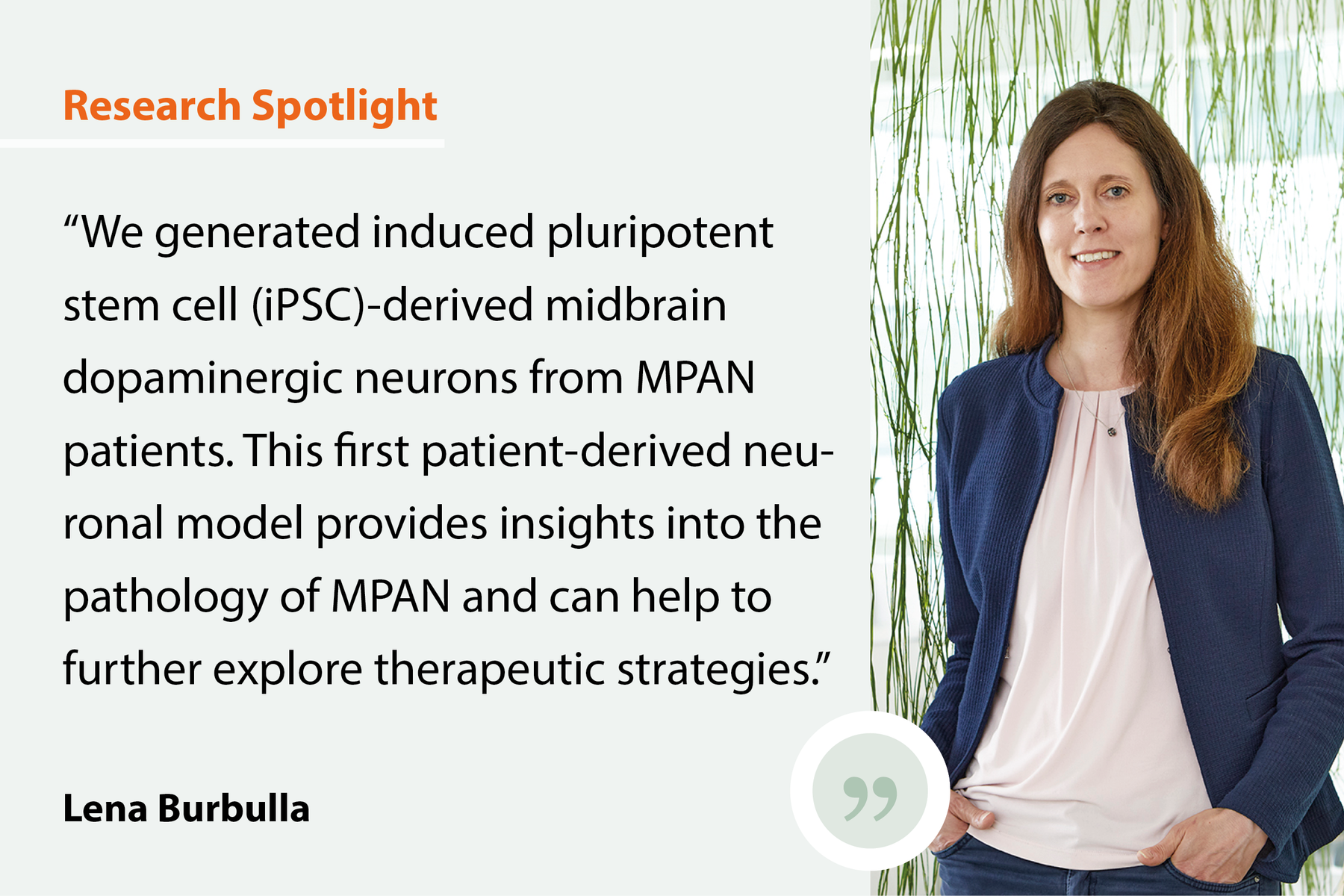
We generated induced pluripotent stem cell (iPSC)-derived midbrain dopaminergic neurons from MPAN patients and examined ultrastructural and biochemical markers of pathology.
Our findings
We found that these neurons displayed the key features of the disease: including α-synuclein and iron accumulation, axonal swellings, and membrane disruption. In addition, levels of the major histocompatibility complex class I (MHC-I), linked to cellular stress and neurodegenerative processes, were elevated in MPAN patient neurons. Treatment with acetyl-leucine, reported to have disease-modifying effects in prodromal PD, decreased these levels.
The implications
A patient-derived neuronal model provides insights into the pathology of MPAN and can help to further explore therapeutic strategies.
Creating SyNergies
The study was led by our member Lena Burbulla and included our members Stefan Lichtenthaler and Martina Schifferer. Our project heavily benefited from the integration of expertise and technologies contributed by several key collaborators. In particular, Martina Schifferer provided cutting-edge electron microscopy technology that was instrumental for our ultrastructural analyses. The proteomics component, which formed a central part of our study, was led by Stefan Lichtenthaler and his team.

